
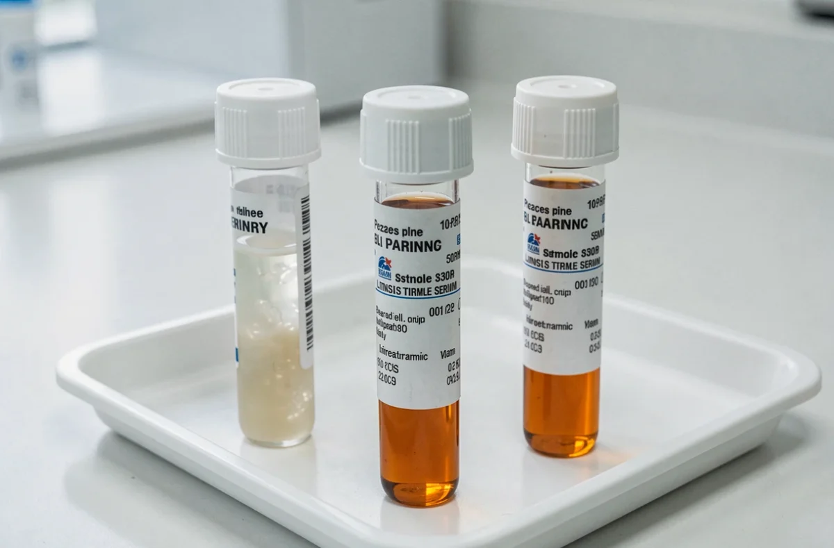
During a lipid panel, the first line of the report often describes the appearance of the serum. This blood fluid, separated from its cells and clotting proteins, is normally clear and pale yellow. After a high-fat meal, it takes on a milky, pearlescent appearance that the laboratory describes as opalescent. This cloudiness indicates the presence of lipid particles suspended in the serum, large enough to scatter light.
Light scattering and circulating lipids: the optical mechanism at work
Serum opalescence is not a measurement. It is a physical phenomenon in which light is scattered, much like a lighthouse beam in fog. When particles of a certain size are suspended in a liquid, they deflect light rays and make the fluid appear cloudy.
In blood serum, the particles responsible are mainly chylomicrons. Produced by the small intestine after dietary fats are digested, these lipoproteins are the largest circulating lipid particles. Their size allows them to scatter visible light, giving the serum its characteristic pearlescent appearance.
One point that is often misunderstood: a cloudy appearance does not reveal the exact triglyceride concentration. The lipemia index measured by automated analyzers depends on the instrument and the laboratory method. Two serum samples that look similar to the naked eye may have different triglyceride levels. Visual assessment is therefore a qualitative indicator, not a quantitative one.
To better understand why serum looks opalescent after a high-fat meal, it is important to distinguish this temporary cloudiness from chronic lipemia, which has a different underlying mechanism.
Post-meal triglycerides: why serum becomes cloudy after eating
Dietary fat absorption follows a specific pathway. Fatty acids and glycerol produced during digestion are taken up by enterocytes in the small intestine, reassembled into triglycerides, and then packaged into chylomicrons. These particles enter the bloodstream via the thoracic duct and lymphatic system, not directly through the portal vein.
This pathway explains the delay. Serum does not become opalescent immediately after a meal. It usually happens a few hours after eating fat, when the concentration of chylomicrons in the blood reaches its peak.
Lipoprotein lipase, an enzyme located on the surface of blood capillaries, then gradually breaks down these chylomicrons by taking up their triglycerides for storage or use as an energy source. The serum becomes clear again once the chylomicrons have been cleared from the bloodstream. This clearance takes several hours, which explains why laboratories traditionally required patients to fast before a blood test.
Lipid panels and fasting: a requirement that is becoming less common
The joint consensus statement from the European Atherosclerosis Society (EAS) and the European Federation of Clinical Chemistry and Laboratory Medicine (EFLM), published in 2016, changed the approach. According to these recommendations, most lipid profiles can be assessed without prior fasting.
The reasoning is straightforward: in everyday life, we spend most of the day in a post-meal state. A non-fasting test therefore reflects typical metabolic conditions. The rise in triglycerides after an ordinary meal is generally modest and does not compromise the interpretation of LDL or HDL cholesterol.
However, the consensus distinguishes between three situations:
- Mild opalescence after a typical meal, which does not affect the reliability of routine tests (total cholesterol, LDL, HDL)
- Marked lipemia (serum that is distinctly milky), which requires cautious interpretation and can distort some results, particularly indirect measurements of sodium or other electrolytes
- Persistent opalescence despite fasting, which may point to an underlying condition (familial hypertriglyceridemia, poorly controlled diabetes, hypothyroidism)
The effect of lipemia on test results is not uniform. The same lipemic blood sample may be suitable for some tests and unusable for others, depending on the laboratory’s analytical method. Direct electrodes for measuring sodium, for example, avoid the artifact caused by the reduced aqueous fraction of lipemic serum.

Persistent serum opalescence: when lipemia is no longer routine
Cloudy serum after a large meal is not in itself a cause for concern. The situation is different when opalescence persists in a sample taken after more than twelve hours of fasting.
Several conditions can keep the level of lipid particles in the bloodstream elevated:
- Primary (familial) hypertriglyceridemia, caused by a genetic defect in the metabolism of triglyceride-rich lipoproteins
- Poorly controlled type 2 diabetes, in which insulin resistance promotes excess hepatic production of VLDL
- Untreated hypothyroidism, which slows the clearance of circulating lipids
- Certain medications (corticosteroids, atypical antipsychotics) that alter lipid metabolism
The cardiovascular risk associated with high triglycerides was long considered secondary to LDL cholesterol. Recent evidence has added nuance to this view. Chronically elevated triglycerides increase cardiovascular risk independently of LDL levels. Regularly opalescent serum in a fasting sample therefore warrants further investigation, including lipoprotein lipase testing or thyroid function tests, depending on the clinical context.
Protein particles: a less familiar cause of cloudiness
Lipids are not the only possible cause of cloudy serum. Abnormal protein particles, such as cryoglobulins or excess immunoglobulins (monoclonal gammopathies), can also scatter light and make serum appear opalescent. These cases are rare, but they are a reminder that cloudiness does not automatically mean excess fat.
When serum is opalescent, the medical laboratory scientist has tools to distinguish a lipid-related cause from a protein-related one. The lipemia index measured by the analyzer, together with triglyceride testing, can quickly guide the diagnosis. Cloudy serum with normal triglycerides suggests looking beyond dietary fats. The visual appearance of serum is a warning sign, not a diagnosis; it is up to the doctor to determine what it means in the context of the patient’s clinical picture.